
“…what drives me is just a very simple thing -- the excitement one gets of understanding something first.”1
−Richard Van Duyne, American Physical Chemist (1945-2019)
It would be an understatement to say that surface-enhanced Raman spectroscopy (also referred to as surface-enhanced Raman scattering or SERS) has evolved into a field of its own. Searching “SERS” online reveals tens of thousands of articles, showing that, since the accidental discovery of SERS in the 1970s, the technique had a tremendous impact on research in chemistry. At the core of SERS is the phenomenon where Raman scattering by molecules is greatly enhanced on rough metal surfaces or nanostructures. The enhancement is so significant, as much as1011, that it can be used to detect single molecules, and while the mechanism behind it is not entirely understood, SERS has exploded in utility owing to the ultrasensitive and broad nature of the method, making it useful in diverse applications from medicine to food safety to environmental monitoring and even fine art preservation.
The story of SERS began, as with many important scientific discoveries, with the observation of an unexpected phenomenon. In 1974, Martin Fleischmann, Patrick Hendra, and Jim McQuillan at the University of Southampton, UK, reported the observation of an usually intense Raman signal from pyridine adsorbed on an electrochemically-roughened silver electrode in an aqueous solution.2 Richard Van Duyne at Northwestern University, USA, who was later credited to have first used the term SERS in 1979, described the results from Fleischmann and McQuillan’s publication as “eye-popping” at the time.3 He had been working on the problem of low signal output from Raman interactions with pyridine and had an opportunity to visit Fleischmann’s laboratory shortly after the observation had been made. Van Duyne discussed with Fleischmann and McQuillan possible explanations for the increase in Raman signal, which McQuillan contributed to increased surface area. With this observation of increased Raman signal and the lack of understanding of the mechanism behind it, laboratories around the world, including Van Duyne’s, were eager to begin their own investigations to figure out what was happening.
In 1977, Van Duyne and his graduate student, David Jeanmaire, carried out a series of experiments and found that the signal intensity per molecule on a surface compared to in free solution had an enhancement of 105–106, a result that could not be explained by surface area alone.4 In their article, they concluded that the increase in signal intensity arose from an enhanced static electric field in the electrochemical double layer.4 A year later, Martin Moskovits at University of Toronto, Canada, took the idea a step further by proposing a localized surface plasmon resonance mechanism to explain the increase in observed signal. This enhancement subsequently became known as “electromagnetic enhancement.”5 Moskovits would later say that the “…the electromagnetic theory should more correctly be called plasmonic enhancement,” in recognition of the important role of surface plasmons in the process.6 Independent of Van Duyne’s work in 1977, M. Grant Albrecht and J. Alan Creighton at the University of Kent, UK, observed similar increases in Raman intensity,7 and two years later, reached a similar conclusion as Moskovits on the importance of plasmons.8 In 1978, Van Duyne and his longtime collaborator, George Schatz, also at Northwestern University, USA, published a paper providing a theoretical description of the intriguing Raman signal enhancements (Figure 1).9 We recommend refs 10 and 11 for a more detailed and comprehensive review on the discovery, development, and application of SERS.
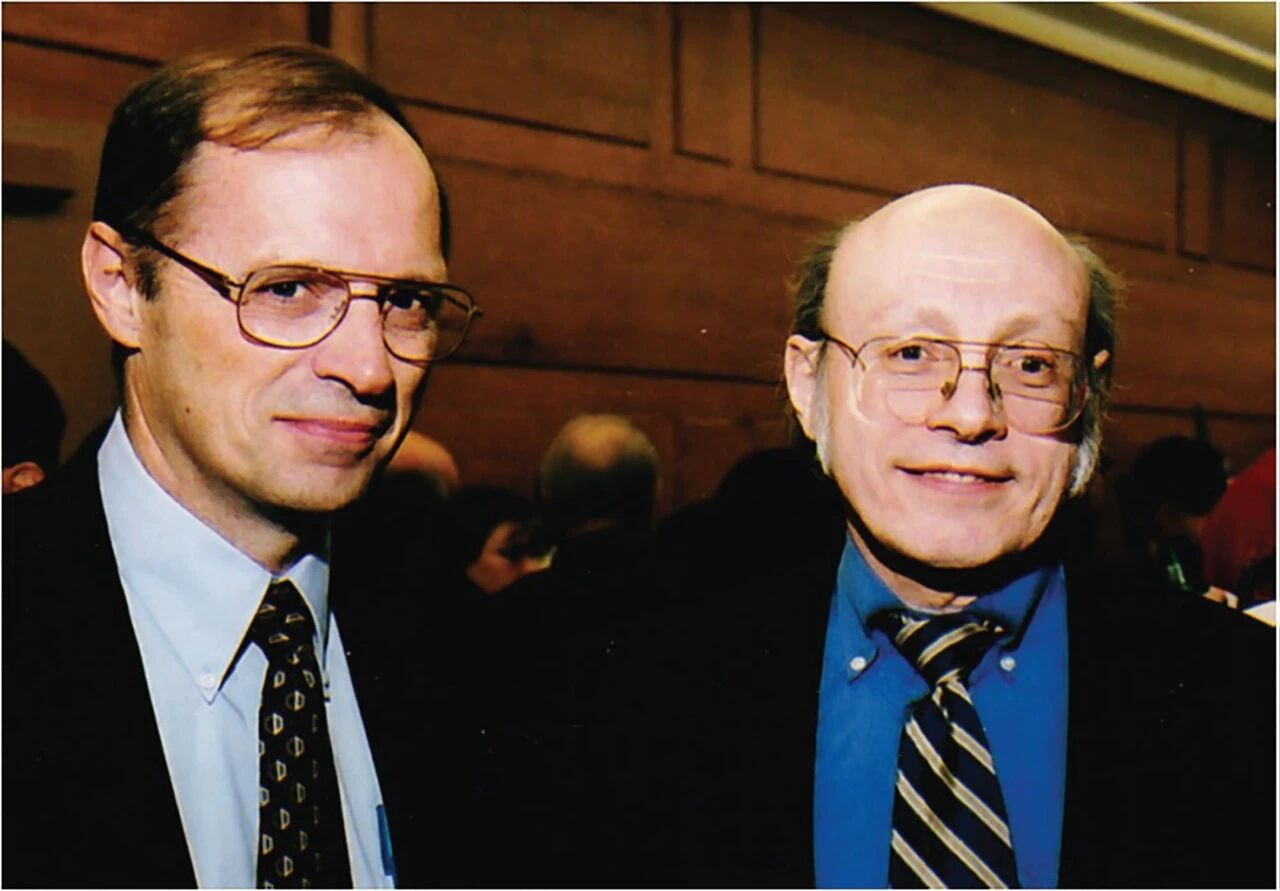
Figure 1 | Geroge Schatz (L) and Richard “Rick” Van Duyne (R), longtime collaborators who played an instrumental role in understanding the mechanism of SERS. [Photo courtesy of the Northwestern University Department of Chemistry]
The exact mechanism of SERS continues to be discussed; however there is scientific consensus that the observed increase in Raman intensity in SERS is dominated by electromagnetic enhancement, which can account for a 104 to 108 increase in signal intensity.12Another contribution, called “chemical enhancement,” which describes the chemical interaction between molecules chemisorbed on the SERS-active surface and the surface itself, can also be present, but provides a significantly smaller boost in intensity, typically 101 to 102 magnitude.12
Since the first discovery of SERS, the technique has been explored under a variety of conditions with different surfaces (metal and nonmetal) and target molecules. Some capabilities of SERS have proven to be unique strengths of the method and are highlighted primarily in three domains: (1) single-molecule detection, (2) in situ monitoring of electrochemical interfaces, and (3) noninvasive deep tissue detection.
Single-molecule detection: Examples for single-molecule detection include experiments from Nie and Emory13 and Kneipp et al.14 who independently reported single-molecule SERS phenomena on silver nanoparticle colloids, a breakthrough that pioneered a novel spectroscopic method beyond single-molecule fluorescence. They demonstrated that SERS was capable of providing high-energy-resolution molecular vibrational fingerprints while circumventing common photobleaching issues encountered in fluorescence techniques.
In situ monitoring of electrochemical interfaces: Owing to the insensitivity of Raman spectroscopy to interference from aqueous solutions, SERS has become an essential tool for investigating electrochemical processes at solid–liquid interfaces. For example, Ren and coworkers15 developed an STM-based electrochemical Tip-enhanced Raman spectroscopy (TERS) technique, overcoming optical distortion in liquid phases through a horizontal incidence light path design, and achieved nanoscale-resolved observation of molecular configuration transitions on Au(111) surfaces. To study supported nanocatalysts, Fu, Chen, Li, and coworkers16developed shell-isolated nanoparticle-enhanced Raman spectroscopy (SHINERS) by assembling catalyst nanoparticles onto the surface of shell-isolated nanoparticles (SHINs) to form a Au@SiO2@catalyst structure. Leveraging plasmonic coupling, this approach increased the enhancement factor by 2–3 orders of magnitude, enabling in-situ studies of catalytic reactions.
Noninvasive deep tissue detection: Matousek and coworkers17 combined spatially offset Raman spectroscopy with SERS to develop surface-enhanced spatially offset Raman spectroscopy (SESORS) that extends penetration depth to millimeter and even centimeter scales, opening new avenues for rapid, label-free, and noninvasive in vivo diagnostics.
An important question that is being addressed is what substrates other than Au and Ag can be used to observe the SERS phenomenon. Both Au and Ag have completely filled d orbitals and a single ‘free’ s electron which creates the surface-localized electric fields. The line of investigation into broadening candidate substrates has led to many significant breakthroughs, one of which was uncovered by Tian and coworkers.18By combining confocal Raman spectroscopy with electrochemical roughening procedures, they obtained SERS signals on transition metal surfaces including Pt, Fe, Ru, Rh, Co, Ni, and Pd, challenging the long-held notion that SERS was only applicable to free-electron metals.18 Furthermore, they developed SHINERS, as mentioned above, utilizing a core-shell structure of Au/Ag cores coated with an ultrathin (1–5 nm) SiO2 shell. The Au core provides a strong electromagnetic field, while the inert SiO2 shell isolates the metal core from direct contact with the probe, enabling noncontactdetection and overcoming the limitations of conventional SERS regarding material types and surface morphologies.19The search for different substrates and potential uses continues. “If we can learn to tune a substrate to any excitation wavelength from say ∼320 nm—which is the cut-off point for silver—to 1200 nm, the game will change,” Van Duyne suggested.3
With all the potential SERS holds, there is no doubt that many more exciting discoveries are on the horizon. We highlight one particular application in the present issue in a Mini Review by Yukuhiro Ozaki, Weidong Ruan, and coworkers on detecting bacterial biological warfare agents. In addition, this month’s issue contains three more Mini Reviews discussing: (1) the impact of combustion on the atmosphere and climate by Katharina Kohse-Höinghaus et al., (2) ultrafast synthesis of zeolite by Zhendong Liu, Javier García-Martínez, et al., and (3) electrochemical ammonia synthesis by Pengfei Wei, Guoxiong Wang, et al. The issue also contains four Communications and 32 Research Articles, covering C–H activation, cyclophanes, iron-catalyzed skeletal editing, total synthesis, metal–organic frameworks, photocatalytic hydrogen production, quantum dynamics, lithium–sulfur battery, shape memory polymers, organotin-oxo clusters for lithography, enzyme-activatable fluorescent probes, plastic scintillators, and much more. A summary of each article in the issue is provided here.
It is fascinating to reflect upon how the discovery of a single technique like SERS can have such a profound impact on a field and careers of so many. Van Duyne, as with others engaged in the scientific enterprise, was a kind and engaged mentor, sharing his excitement of discovery and propagating further growth of students and the research. In a reflection of Van Duyne’s career by Schatz, he stated that, “…he [Van Duyne] was always excited by the quest for scientific advance, and he was excellent at conveying that excitement to those around him.”20 Even up to the years leading up to his death in 2019, Van Duyne could be seen in office working to untangle the scientific challenges that lie ahead.20
The story of SERS over the past 50 years is a superb example of how science builds up over decades through combined dedication, intellect, and contributions of countless talented and thoughtful people. Scientific results do not arise solely from a single individual and are not confined within a certain decade of progress. The scientific discoveries that can be realized now only exist because of years of thought, planning, and experimentation by great scientists that came before us and who immersed themselves in, as Van Duyne remarked in an interview in 2013, “…the excitement one gets of understanding something first.”1 All of us performing scientific research at any level have the privilege of being a member of this elite group of pioneers who choose to follow their insatiable curiosity and use their time and abilities to do the detailed, tedious, and rewarding work of building a global scientific legacy.
Prof. Dr. Xi Zhang
Editor-in-Chief
E-mail: xi@tsinghua.edu.cn
Dr. Donna J. Minton
Director of Publications, CCS
E-mail: donna.minton@chinesechemsoc.org
References:
1. Van Duyne, R. P.Professor Richard Van Duyne.International Institute for Nanotechnology YouTube, Feb. 6, 2013. https://www.youtube.com/watch?v=u2u49kzqsq4
2. Fleischmann, M.; Hendra, P. J.; McQuillan, A. J.Raman Spectra of Pyridine Adsorbed at a Silver Electrode.Chem.Phys. Lett.1974,26, 163–166.
3. Martin, D.SERS and the Rise of the Raman EmpireChemistryWorldhttps://www.chemistryworld.com/features/sers-and-the-rise-of-the-raman-empire/9264.article. (Accessed March 13, 2026).
4. Jeanmaire, D. L. and Van Duyne, R. P.Resonance Raman Spectroelectrochemistry. 3. Tunable Dye Laser Excitation Spectroscopy of the Lowest 2B1u Excited State of the Tetracyanoquinodimethane Anion Radical.J. Electroanal. Chem.1977,84, 1–20.
5. Moskovits, M.Surface Roughness and the Enhanced Intensity of Raman Scattering by Molecules Adsorbed on MetalsJ. Chem. Phys.1978,69, 4159-4161.
6. Moskovits, M. and Piorek, B. D.Brief History of Surface-Enhanced Raman Spectroscopy and the Localized Surface Plasmon Dedicated to the Memory of Richard Van Duyne (1945–2019)J. Raman Spectrosc.,2021,52, 279–284.
7. Albrecht, M. G. and Creighton, J. A.Anomalously Intense Raman Spectra of Pyridine at a Silver Electrode.J. Am. Chem. Soc.1977,99, 5215–5217.
8. CreightonJ. A.; BlatchfordC. G.; Albrecht M. G.Plasma Resonance Enhancement of Raman Scattering by Pyridine Adsorbed on Silver or Gold Sol Particles of Size Comparable to the Excitation Wavelength.J. Chem. Soc., Faraday Trans. 2,1979, 75, 790–798.
9. KingF. W.; Van DuyneR. P.; SchatzG. C.,Theory of Raman Scattering by Molecules Adsorbed on Electrode Surfaces.J. Chem. Phys.1978,69, 4472–4481.
10. Yi, J.; You, E-M; Ren, H.; Wu, D-Y; Liu, G-K; Yang, Z-L.; Zhang, H.; Gu, Y.; Wang, Y-H; Wang, X., et al.Surface-enhanced Raman Spectroscopy: A Half Century Historical Perspective.Chem. Soc. Rev.,2025,54, 1453-1551
11. Langer, J; de Aberasturi, D. J.; Aizpurua, J.; Alvarez-Puebla, R. A.; Auguié, B; Baumberg, J. J.; Bazan, G. C.; Bell, S. E. J.; Boisen, A.; et al.Present and Future of Surface-Enhanced Raman Scattering.ACS Nano2020,14, 28−117.
12. Stiles, P. L.; Dieringer, J. A.; Shah, N. C.; Van Duyne, R. P.Surface-Enhanced Raman Spectroscopy.Annual Rev. Anal. Chem.2008,1, 601–626.
13. Nie, S. and Emory, S. R.Probing Single Molecules and Single Nanoparticles by Surface-Enhanced Raman Scattering.Science,1997,275, 1102–1106.
14. Kneipp, K.; Wang, Y.; Kneipp, H.; Perelman, L. T.; Itzkan, I.; Dasari, R. R.; Feld, M. S.Single Molecule Detection Using Surface-Enhanced Raman Scattering (SERS).Phys.Rev. Lett.,1997,78, 1667–1670.
15. Zeng, Z.-C.; Huang, S.-C.; Wu, D.-Y.; Meng, L.-Y.; Li, M.-H.; Huang, T.-X.; Zhong, J.-H.; Wang, X.; Yang, Z.-L.; Ren, B.Electrochemical Tip-Enhanced Raman Spectroscopy.J. Am.Chem. Soc.,2015,137, 11928–11931.
16. Zhang, H.; Wang, C.; Sun, H.-L.; Fu, G.; Chen, S.; Zhang, Y.-J.; Chen, B.-H.; Anema, J. R.; Yang, Z.-L.; Li, J.-F.; Tian, Z.-Q.In Situ Dynamic Tracking of Heterogeneous Nanocatalytic Processes by Shell-isolated Nanoparticle-enhanced Raman Spectroscopy.Nat. Commun.,2017,8, 15447.
17. Stone, N.; Faulds, K.; Graham, D.; Matousek,P. Prospects of Deep Raman Spectroscopy for Noninvasive Detection of Conjugated Surface Enhanced Resonance Raman Scattering Nanoparticles Buried within 25 mm of Mammalian Tissue.Anal. Chem.,2010,82, 3969–3973.
18. Ren, B.; Huang, Q. J.; Cai, W. B.; Mao, B. W.; Liu, F. M.; Tian, Z.-Q.Surface Raman Spectra of Pyridine and Hydrogen on Bare Platinum and Nickel Electrodes.J. Electroanal. Chem.,1996,415, 175–178.
19. Li, J.-F.; Huang, Y. F.; Ding, Y.; Yang, Z.-L.; Li, S. B.; Zhou, X.-S.; Fan, F.-R.; Zhang, W.; Zhou, Z.-Y.; Wu, D.-Y.; Ren, B.; Wang, Z. L.; Tian, Z.-Q.Shell-isolated Nanoparticle-enhanced Raman Spectroscopy.Nature,2010,464, 392–395.
20. Schatz, G. C.SERS and the Scientific Career of Richard P. Van Duyne (1945–2019).J. Raman Spectrosc.2021,52, 268–278.
Link to the original text:
https://doi.org/10.31635/ccschem.026.202600324ed1
